
Dejan Galič, MD, FEBU, FECSM
urology specialist
He deals with general urology, especially urological oncology and prostate problems, as well as sexual medicine.


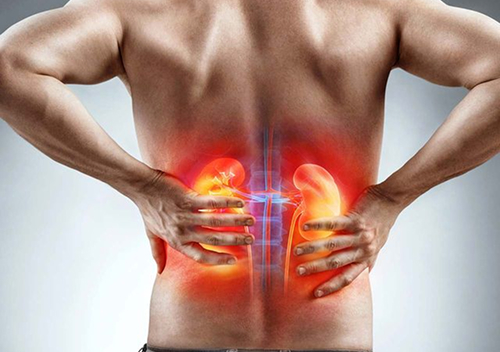
Urinary stones (urolithiasis), commonly called kidney stones, form when urine is too concentrated with certain minerals and other substances, which begin to separate out as tiny crystals. The crystals can bind together and gradually grow into a stone. Stones can be found in the kidneys, ureter or bladder, and less often in other parts of the urinary tract. By composition, calcium stones are the most common (80%), mainly calcium oxalate and calcium phosphate, but other types also occur, such as uric acid stones, struvite (infection) stones, cystine stones and many others.
Urolithiasis is a common urinary tract disease whose incidence is rising in many countries. The risk of stone formation is linked to genetic, metabolic, dietary and environmental factors. Important risk factors include insufficient fluid intake, concentrated urine, a diet high in salt and animal protein, excess body weight, metabolic syndrome, certain metabolic diseases, urinary tract infections, certain medicines and a family or genetic predisposition. Previous urolithiasis is also an important risk factor, as stones often recur.
Urolithiasis can cause no symptoms at all, or it can cause typical problems, renal colic, especially when a stone moves into the ureter and partly or completely blocks the flow of urine. Renal colic is a sudden, very strong cramping pain in the flank that may spread to the lower abdomen, groin or genitals. It is often accompanied by blood in the urine, nausea and vomiting, and if the stone is in the lower part of the ureter there may also be more frequent and urgent urination and burning when urinating. Fever, chills or feeling generally unwell with a blocked urinary tract may indicate an infection and require prompt urological care.
Based on a typical history and clinical picture, the urologist suspects a stone in the urinary tract. Further diagnostics then aim to confirm the stone, determine its size and position and assess any urinary obstruction and infection. Urine tests and, if needed, blood tests are performed: in the urine we look for haematuria, crystals and signs of infection, and in the blood we check kidney function and other parameters depending on the clinical situation.
Urinary tract ultrasound is often the first imaging test, as it can show kidney stones, dilation of the collecting system and some ureteral stones. For a more precise assessment, a non-contrast abdominal CT is usually the most suitable, as it reliably shows the size, location, number and density of stones and the degree of obstruction. Other imaging methods (e.g. intravenous urography) are used in selected patients.
The treatment of kidney stones depends on the size, position and composition of the stone, the symptoms and any urinary obstruction or infection. If the ureter is blocked and there is an infection, it is a urological emergency requiring an immediate visit to an emergency urology department and urgent drainage of the urinary tract with a JJ stent or nephrostomy; the stone itself is usually removed once the infection has settled. Small (up to 10 mm), asymptomatic kidney stones that do not cause obstruction, infection or impaired kidney function are often monitored, as most of them pass spontaneously; to help them pass, patients receive α-blockers and pain relief (NSAIDs).
For stones larger than 10 mm, the chance of spontaneous passage is small, so active treatment is recommended:
Kidney stones are best prevented by drinking enough fluids, eating a balanced diet, reducing salt and eating animal protein in moderation. In recurrent urolithiasis, analysis of the stone composition, a metabolic evaluation and a 24-hour urine analysis are important to identify individual risk factors and plan targeted preventive measures.


Monday - Thursday
Friday
Saturday - Sunday
08:00 - 19:00
08:00 - 16:00
Closed
Copyright © 2025 MD medicina | All Rights Reserved | Quality and safety | Privacy Policy
 English
  Slovenian
  German
  Italian
  Albanian